
Figure 5.
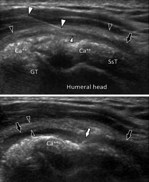
Lateral approach. Oblique coronal US scans (see fig. 1, the right image). Above: large calcifications (Ca + +) of the supraspinatus tendon (SsT). Poorly defined calcifications, significant wall thickening (black arrowheads) and subacromial-subdeltoid bursal fluid collection (arrow) indicate a calcium migratory syndrome (from the tendon to the bursa). The needle (large white arrowheads) was positioned under careful real-time US guidance to avoid damaging the tendon calcifications. The tip (small white arrowhead) is inside the subacromial-subdeltoid bursa. Below: After completion of corticosteroid injection the bursa is distended by the drug (black arrows); note the presence of small air bubbles inside the subacromial-subdeltoid bursa (white arrow).