
Figure 12.
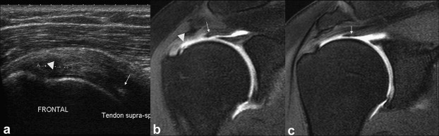
Partial-thickness tear of the deep surface of the supraspinatus tendon. Coronal US scan (a) and arthro-MRI (b, c). The deep fibers of the tendon are disinserted and considerably retracted (arrows) causing a US pitfall. The US image shows some anomalies of the deep surface (arrowheads) at the insertion of the tendon, but does not clearly show the retracted string almost at the base of the humeral head (arrows).