
Figure 1.
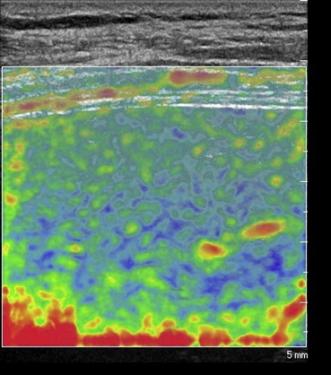
Elastographic image obtained during an intercostal scan performed with a linear transducer. It provides real-time colorimetric representation of liver stiffness: blue indicating the stiffer areas of the parenchyma and red/green those that are softer and more compliant.