
Fig. 1.
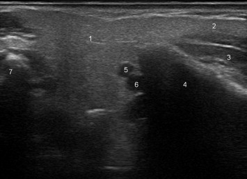
Axial scan of the parotid gland showing the superficial lobe (1), the masseter extension of the gland (2), the masseter muscle (3), the ramus of the mandible (4),the retromandibular vein (5), the external carotid artery (6), and the apex of the mastoid process (7).