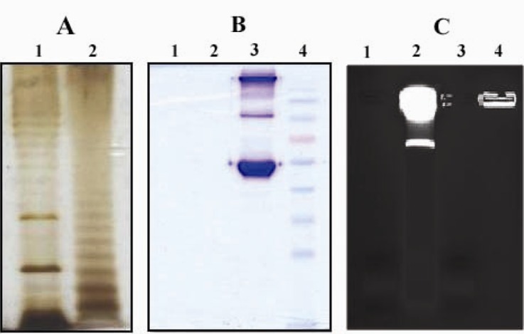
Figure 1.
Silver, coomassie blue and ethidum bromide stainings of purified LPS
LPS from E coli (Lanes 1A and 1B) and S.typhi (lanes 2A and 2B) was purified by modified hot phenol-water extraction method and fractionated by SDS-PAGE electrophoresis followed by silver (A) or commassie blue staining (B). Ladder pattern of LPS banding which is charasteristic of smooth gram negative bacteria is seen (A). The absence of band in commassie blue staining as shown in B indicates no contamination of purified LPS with bacterial proteins. Lane 3B: Human IgG and BSA, Lane 4B: Molecular weight marker. Residual nucleic acid contamination in purified LPS products was traced by eithidium bromide staining (C). Absence of band in LPS from E.coli (Lane 1C) and S.typhi (Lane 3C) shows no contamination with nucleic acids in purified LPS products. Lane 2C and 4C: Whole E. coli and S.typhi, respectively

