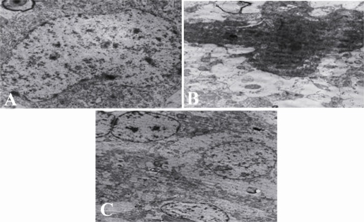
Figure 3.
Electron micrograph of cortical neurons from intact, control, and WK pre-treated groups. Normal ultrastructure is visible in intact neurons. Seizure results in severe degenerative changes in cortical neurons (part B). Cortical neurons from the WK pre-treated group show some degenerative changes, but the whole ultrastructure was maintained (part C). Magnification in (A) and (B) is 8900 × and in (C) is 3900

