
Figure 10.
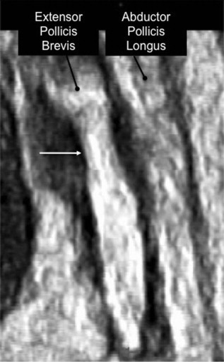
Three-dimensional sonographic appearance of de Quervain tenosynovitis, type II, allows reconstruction in the coronal plane. Note constrictive effects of the retinaculum (arrow) on the extensor pollicis brevis tendon, whereas the abductor pollicis longus tendon has a normal appearance with parallel margins.