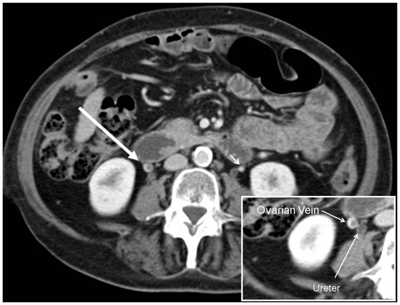
Figure 1.
69 year old woman with metastatic pancreatic cancer and incidentally identified ovarian vein thrombosis complicated by pulmonary embolism. Axial contrast enhanced CT (Siemens Sensation 64, 120 kVp, 325 mAs, 0.75 mm slice thickness, 100 ml Omnipaque 320, portal venous phase) demonstrating a filling defect in right ovarian vein as central hypodensity surrounded by dense contrast (long white arrow), consistent with thrombus. Magnification image insert with ovarian vein thrombus and ureter marked. Note the right ovarian vein is medial to right kidney and anterior to right ureter. The left ovarian vein marked with short arrow demonstrating homogenous opacification without thrombus contrary to right ovarian vein.

