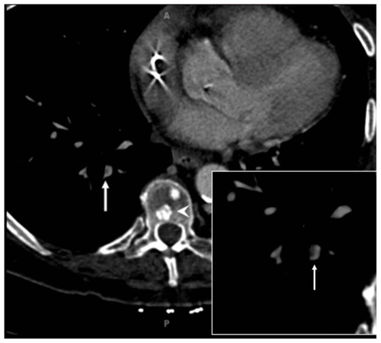
Figure 3.
69 year old woman with metastatic pancreatic cancer and incidentally identified ovarian vein thrombosis complicated by pulmonary embolism. Thin section axial contrast enhanced CT (Siemens Sensation 64, 120 kVp, 325 mAs, 0.75 mm slice thickness, 100 ml Omnipaque 320, arterial phase) in soft tissue window demonstrating subtle filling defect (white arrows) in right lower lobe pulmonary arterial branch, consistent with pulmonary embolism. Magnification view insert demonstrates thrombus as focal hypodensity within vessel (arrow). Sclerotic osseous metastases are denoted by white arrowhead within thoracic vertebral body. Mediport catheter tip artifact is seen in the right atrium (not marked).

