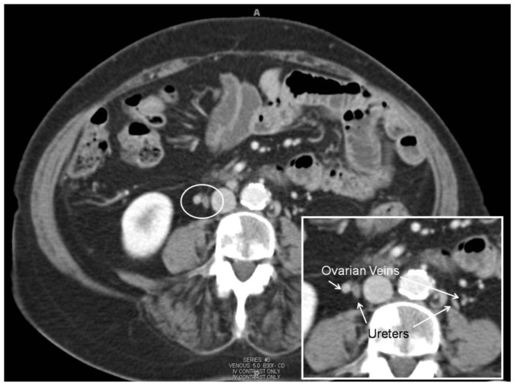
Figure 6.
69 year old woman with metastatic pancreatic cancer and incidentally identified ovarian vein thrombosis complicated by pulmonary embolism. 3 month follow-up CT (Siemens Sensation 64, 120 kVp, 350 mAs, 5 mm slice thickness, 100 ml Omnipaque 320, venous phase) after anticoagulation demonstrating resolution of right ovarian vein filling defect (figure 1a) which now demonstrates well opacified, normal caliber (3–4 mm), right ovarian vein (white circle). Note the sclerotic metastases through the imaged vertebral body (not marked). Magnification insert view demonstrates well-opacified, normal caliber (3–4 mm), both ovarian veins and ureters marked (white arrows). Note normal caliber ovarian veins are anterior and lateral to ureters (marked).

