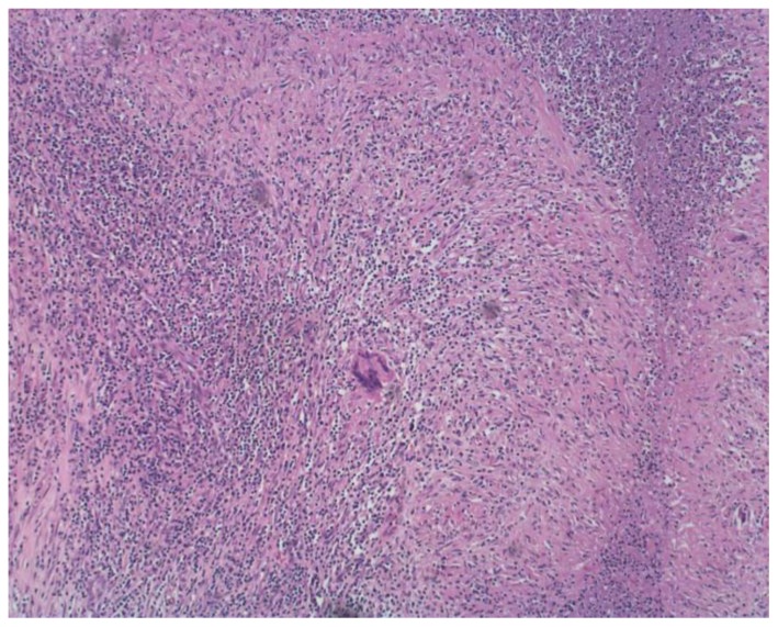
Figure 3.
76-year-old male with left tuberculous epididymo-orchitis. Microscopic pathology, Hematoxylin and Eosin (H&E) stain. Low power view reveals multiple necrotizing granulomas with central areas of necrosis surrounded by collections of epithelioid histiocytes as well as many Langerhans multinucleated giant cells and a chronic inflammatory infiltrate.

