
Figure 1.
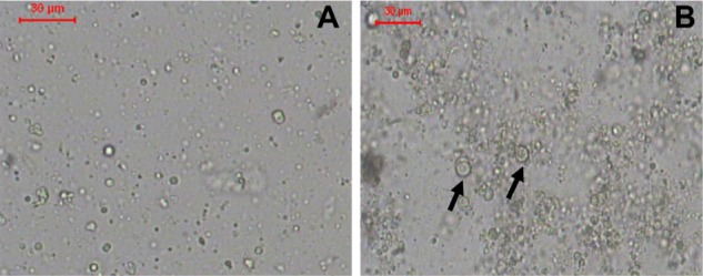
(A and B) Optical photomicrographs of liposomes. (A) F1, prepared by lipid hydration method and (B) F2, prepared by heating method.
Notes: Bar = 30 μm. Arrows point to multilamellar vesicles.
Official websites use .gov
A
.gov website belongs to an official
government organization in the United States.
Secure .gov websites use HTTPS
A lock (
) or https:// means you've safely
connected to the .gov website. Share sensitive
information only on official, secure websites.
(A and B) Optical photomicrographs of liposomes. (A) F1, prepared by lipid hydration method and (B) F2, prepared by heating method.
Notes: Bar = 30 μm. Arrows point to multilamellar vesicles.