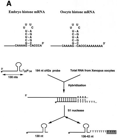
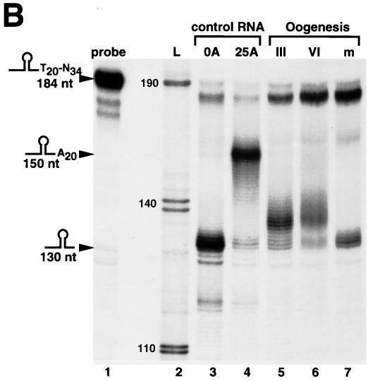
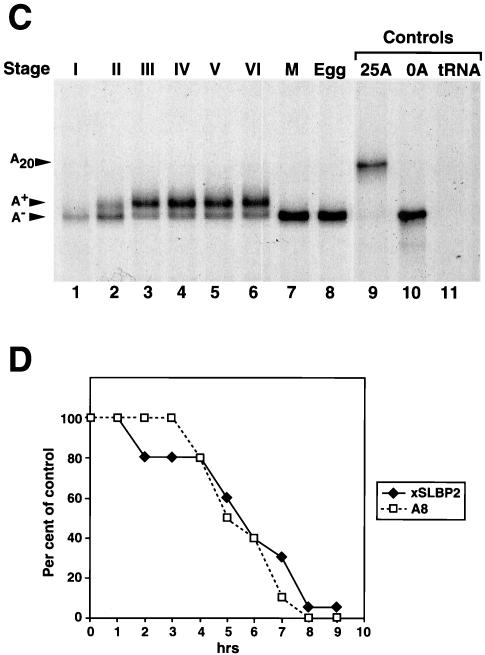
FIG. 1.
Oocyte histone mRNA contains an oligo(A) tail. (A) The structures of the 3′ ends of the egg (left panel) and oocyte (right panel) histone mRNAs are shown. The S1 nuclease protection assay used to determine the length of the poly(A) tail is diagrammed. (B) The S1 nuclease assay (as shown in panel A) was used to map the 3′ end of Xenopus histone H2a mRNA in stage III and stage VI oocytes and matured oocytes. Synthetic RNAs ending in the ACCCA sequence formed by histone pre-mRNA processing (lane 3) or ending in the ACCCA followed by 25 As (lane 4) were incubated with the probe containing 20 Ts after the stem-loop. The reaction mixtures were treated with S1 nuclease, resulting in a fragment of 130 nt resulting from protection of the ACCCA (lane 3) and a 150-nt fragment resulting from protection of the A20 tail (lane 4). For lanes 5 to 7, total cell RNA from one oocyte equivalent of RNA from stage III, stage VI, or matured oocytes (m) was incubated with the probe, the reaction mixtures were treated with S1 nuclease, and the protected fragments were analyzed on a 60-cm-long 8% polyacrylamide-7 M urea gel. Lane 1, a 184-nt probe; lane 2 (L), pUC18 digested with HpaII and labeled with [α-32P]dCTP. The sizes of these fragments are indicated. The fragment at about 175 nt in lanes 1 to 5 is derived from the probe. (C) RNA from one oocyte or egg was analyzed at the indicated stages by S1 nuclease mapping as described in Materials and Methods; the results are presented in lanes 1 to 6. The protected fragments were analyzed on a 10-cm-long 8% polyacrylamide-7 M urea gel. The results are representative of analyses of oocytes from four different frogs. The results of analysis of RNA from oocytes matured by treatment with progesterone are shown in lane 7, and lane 8 shows the results of analysis of RNA from Xenopus eggs. Lanes 9 and 10 show the results of analysis of synthetic RNAs ending in the histone stem-loop and in the stem-loop followed by 25 As, respectively. Lane 11 shows the results of analysis of 10 μg of yeast tRNA. (D) Stage VI oocytes were incubated with progesterone. At 1 h intervals, 20 oocytes were removed and pooled. The oocytes were homogenized in buffer, and RNA was prepared from 80% of the homogenate. The rest of the homogenate (four oocytes) was analyzed by sodium dodecyl sulfate (SDS)-gel electrophoresis, and xSLBP2 was detected by Western blotting. The S1 analysis was done using RNA from four oocytes. The percentages of histone mRNA containing oligo(A) tails and the amounts of xSLBP2 (as determined by Western blotting) are indicated, with 100% representing the levels prior to addition of progesterone. At 7 h 70% of the oocytes had matured (as determined by the appearance of the white spot at the animal pole), and at 8 and 9 h all the selected oocytes had matured. No oocytes had matured at 6 h.


