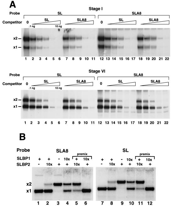
FIG. 3.
xSLBP1 and xSLBP2 bind equally well to the stem-loop and the oligoadenylated stem-loop. (A) Extracts from stage I (top panel) or stage VI (bottom panel) oocytes were incubated with the stem-loop (SL) probe (lanes 1 to 11) or the radiolabeled oligoadenylated stem-loop probe (lanes 12 to 22). Increasing amounts of competitor stem-loop (0.1 to 10 ng; lanes 2 to 6 and lanes 13 to 17) or oligoadenylated stem-loop (0.1 to 10 ng; lanes 7 to 11 and lanes 18 to 22) were mixed with the probe prior to addition of the oocyte lysate. Complexes were resolved by electrophoresis on 8% polyacrylamide gels under native conditions and detected by autoradiography. The positions of the complexes with xSLBP1 (x1) and xSLBP2 (x2) are indicated. Lanes 1 and 12 were analyzed without an added competitor. (B) xSLBP1 and xSLBP2 were synthesized in reticulocyte lysates in the presence of [35S]methionine, and the amounts of each protein were estimated by autoradiography of the in vitro-synthesized proteins. The amounts of xSLBP1 and xSLBP2 required to completely shift the stem-loop probe or the SLA8 probe were determined. Mobility shift assays were performed with (+) or without (−) radiolabeled SLA8 probe (lanes 1 to 6) or the stem-loop probe (lanes 7 to 12). The same amount of xSLBP1 or xSLBP2 was incubated with the stem-loop probe (lanes 1, 3, 7, and 9). After incubation for 5 min at 4°C, a 10-fold excess (10x) of the other xSLBP was added (lanes 2, 4, 8, and 10) and the reaction mixture was incubated for 60 min prior to analysis. For lanes 5, 6, 11, and 12, the two proteins were mixed prior to the addition of the probe (premix).

