Figure 2.
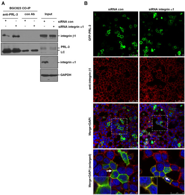
Physical interaction between PRL-3 and integrin β1. (A) Deletion of integrin α1 resulted in enhanced PRL-3-integrin β1 interaction. BGC823 cells were transfected with 100 pmol integrin α1-specific siRNA or control siRNA for 48 hr. 500 μg protein lysates were incubated with PRL-3 antibody or pre-immune mouse IgG (control) for co-immunoprecipitation assay. The immunoprecipitates and 50 μg protein lysates (input) were immunoblotted by integrin β1 and PRL-3 antibodies. Efficiency of intergrin α1 silencing in the input was verified. “LC” is short for light chain of antibody. (B) PRL-3 co-localized with integrin β1 in an integrin α1-independent manner. After being transfected with 100 pmol integrin α1 siRNA and control siRNA for 24 hours, BGC823 cells were transfected with 2 μg GFP-PRL-3, and cultured for another 24 hours, then fixed, blocked, and immunostained with anti-integrin β1 antibody. Localization of GFP-PRL-3 (green) and integrin β1 (red) was detected by a laser confocal microscope. Parts of merged images were enlarged (white rectangles) to show the co-localization between two molecules (yellow). The white arrows labeled regions were further enlarged (insert).

