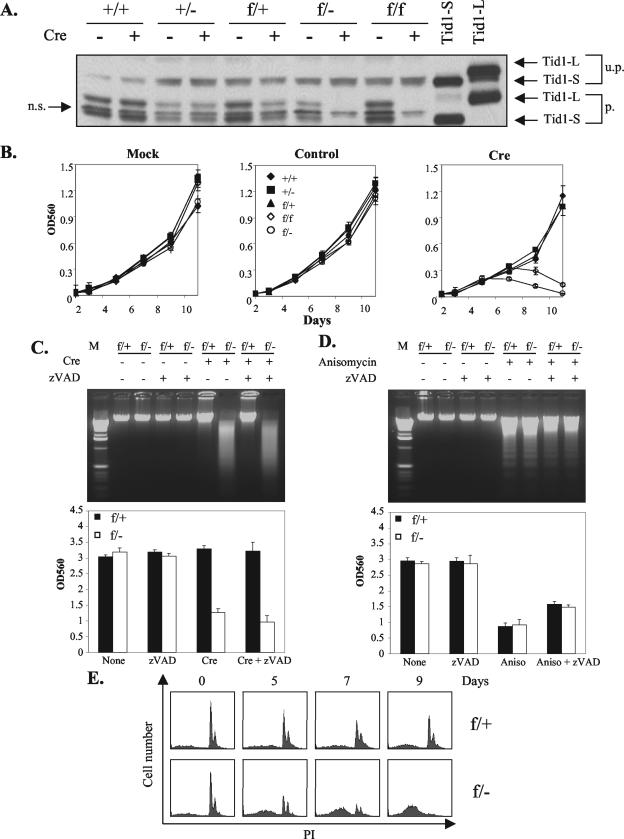
FIG.3.
Tid1 gene product is required for cell survival. (A) MEFs with Tid1+/+, Tid1+/−, Tid1f/+, Tid1f/−, and Tid1f/f genotypes were infected with Ad-Cre (Cre) (+) or uninfected (−) as indicated. After 6 days, total cellular proteins from these cells were collected and separated by SDS-polyacrylamide gel electrophoresis and analyzed by Western blotting with an antibody against mouse Tid1. The recombinant Tid1L and Tid1S proteins generated from 293T cells transfected with expression plasmids encoding Tid1-L or Tid1-S (two splicing isoforms of Tid1) were used as controls. u.p., unprocessed; p., processed; n.s., nonspecific. (B) After infection with nothing (Mock), control adenovirus (control), or Ad-Cre (Cre) as indicated, the above-mentioned MEF cell lines were analyzed for growth and viability by MTT (3-[4,5-dimethylthiazol-2-yl]-2,5-diphenyltetrazolium bromide) assays at the indicated time intervals. OD560, optical density at 560 nm. The error bars indicate standard deviations. (C) Tid1f/+ and Tid1f/− MEFs were treated as for panel A. After 9 days, genomic DNAs were purified from these cells and separated by agarose gel electrophoresis (top). Some MEFs were also treated with 50 μM zVAD (a caspase 3 inhibitor) as indicated. The relative amounts of viable cells for these treated MEFs were also assessed by MTT assays (bottom). (D) Tid1f/+ and Tid1f/− MEFs were treated with 5 μg of anisomycin/ml (50 μM zVAD) as indicated for 18 h, and their genomic DNAs (top) and viabilities (bottom) were analyzed as for panel C. The MTT assays were performed in triplicate and repeated at least three times (the values are means plus standard deviations). (E) Tid1f/+ and Tid1f/− MEFs were infected with Ad-Cre and collected at the indicated time intervals. The cells were fixed and stained with propidium iodide (PI) and analyzed by flow cytometry.

