
Figure 4.
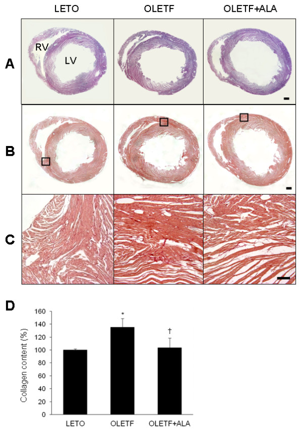
Effect of ALA on cardiac collagen deposition in OLETF rats. Representative micrographs of H&E-stained (A) and Sirius red-stained (B) heart sections from each group. The black lined-box (in B) shows a high-magnification micrograph (C) of Sirius red-stained sections. Scale bar = 1000 μm (200 μm in C). (D) Sircol collagen assay quantifying soluble collagen in the hearts of LETO and OLETF rats with or without ALA. Data are presented as the mean ± SEM. *p < 0.05 vs. LETO rats; †p < 0.05 vs. OLETF rats.