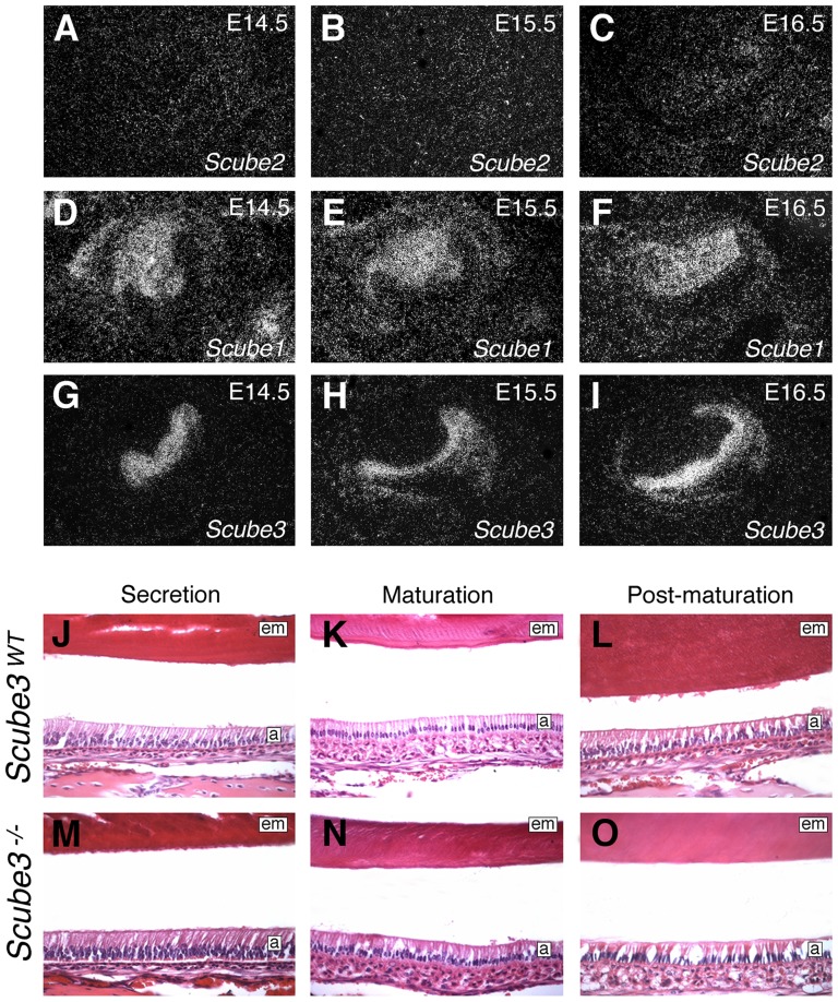
Figure 7. Scube gene function in tooth development.
(A–I) Scube gene expression in the developing first molar at E14.5–16.5 assayed by in situ hybridization on frontal sections. (A–C) Scube2; (D–F) Scube1; (G–I) Scube3. (J–O) Ultra-structural view of incisor tooth development in wild type and Scube3−/− mice at P18. In the incisor, amelogenesis commences apically and proceeds in an coronal direction, demonstrating (J, M) Secretion; (K, N) Maturation; (L, O), Post-maturation. These different stages of amelogenesis all show a characteristically normal morphology in wild-type and Scube3−/− mice. a, ameloblasts; em, enamel matrix.

