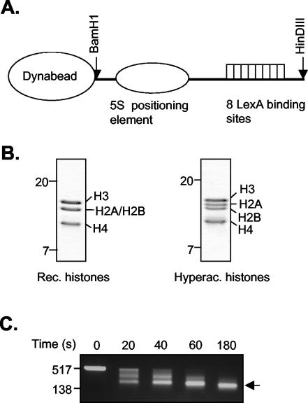
FIG. 3.
Reconstitution and analysis of the nucleosomal template. (A) Schematic representation of the DNA template containing eight LexA binding sites and a 5S nucleosome positioning element. (B) Analysis of purified recombinant (Rec.) Xenopus octamers and hyperacetylated (Hyperac.) core histones purified from HeLa cells on SDS-polyacrylamide (15%) gel electrophoresis gel stained with Coomassie brilliant blue. (C) Partial micrococcal nuclease digestion. Nucleosomal templates were incubated with 10 mU micrococcal nuclease at 37°C for 0, 20, 40, 60, and 180 s. Reactions were stopped by adding 10 mM EGTA. DNA was phenol chloroform extracted, precipitated, and loaded onto a 1.5% agarose gel. DNA size markers are indicated on the left. An arrow indicates mononucleosomal DNA.

