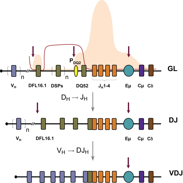
Figure 1. Immunoglobulin heavy chain locus and B cell development.
Schematic of the murine IgH locus showing variable (VH, blue, n represents approximately 150 VH gene segments), diversity (DH, grey, n represents six to nine DSP gene segments), and joining (JH, orange) gene segment [11],[12]. Exons encoding the constant regions of IgM and IgD are indicated as Cμ and Cδ. A promoter 5′ of DQ52, the 3′-most DH gene segment, is indicated by the yellow oval and the intronic enhancer Eμ by a teal oval. Top line shows the germline (GL) configuration with associated histone modifications in B lineage precursors [23]–[25]. Histone H3 and H4 acetylation are shown in orange and presence of heterochromatic H3K9 methylation by the red line. Vertical red arrows represent the tissue-specific DNase I hypersensitive sites in the germline state. Next two lines show sequential stages of VDJ recombination at the IgH locus. DH to JH rearrangement occurs first resulting in a DJH junction and, depending on which DH rearranges, residual upstream unrearranged DHs may be present. VH rearrangement occurs to the DJH junction to generate a VDJH junction; during this process unrearranged DHs are lost from the genome.

