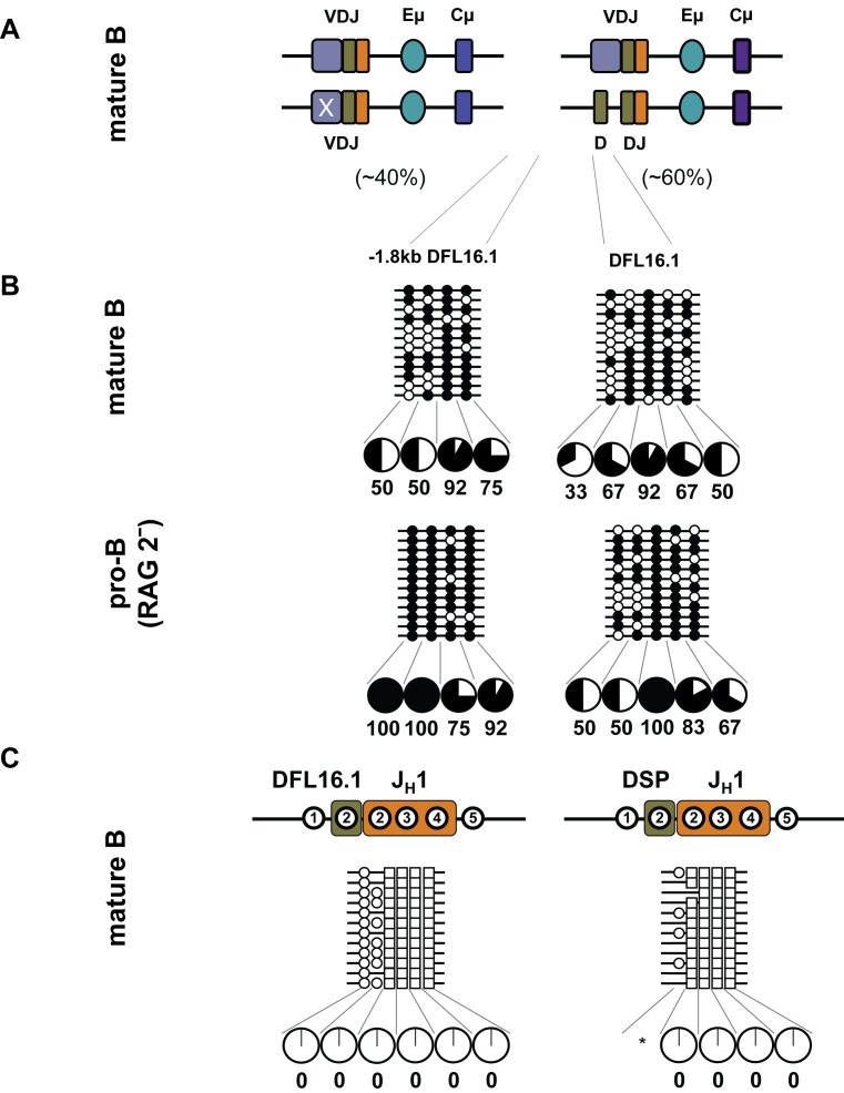
Figure 5. DNA methylation state of unrearranged and DJH recombined alleles in mature B cells.
(A) Mature B cells were purified from spleens of C57BL/6 mice, and the genomic DNA was subjected to bisulfite modification assays. 40% of these cells contain two VDJH recombined alleles and the remainder contains one VDJH and one DJH recombined allele. (B) Amplicons corresponding to unrearranged DFL16.1 gene segment and a region centered 1.3 kb 5′ to DFL16.1 were cloned and sequenced. For comparison, methylation of the same region in pro-B cells derived from RAG2-decificient bone marrow is shown in the bottom panel. Filled and open circles indicate methylated and unmethylated cytosines. Pie charts summarize the percentage of methylated cytosines at each position; data are derived from two independent spleen B cell preparations with two to four mice in each experiment. (C) DJH junctions were amplified from bisulfite modified DNA, followed by cloning and sequencing. Circles and squares represent cytosines from DH and JH1 gene segment, respectively. Filled and open circles, or squares, indicate methylated and unmethylated cytosines, respectively. Numbers within regions marked as DFL16.1, DSP, and JH1 denote CpG dinucleotides corresponding to the configuration at the respective unrearranged gene segments. For example, of the five CpGs at unrearranged DFL16.1, only the first two are retained in DFL16.1/JH1 junctions. Variations in the total number of cytosines are due to imprecise joining during VDJ recombination. Pie charts summarize the percentage of methylated cytosines. The asterisk indicates positions where less than 12 CpGs were observed due to reduced representation caused by junctional diversity.

