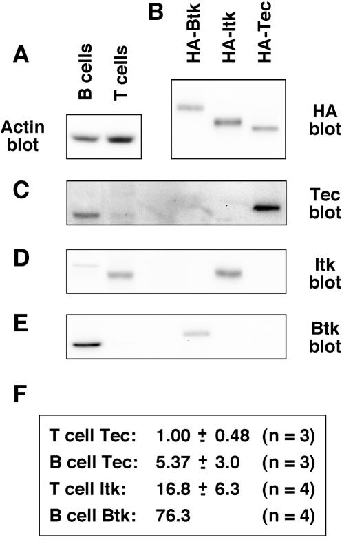
FIG. 1.
Tec expression is relatively low in primary mouse T and B cells, relative to Itk and Btk. (A) B and T lymphocytes were isolated to 90 to 95% purity by MACS from mixed spleen and lymph nodes from 5- to 6-week-old C57BL/6 mice. Cells were lysed, and approximately 5 × 10⁶ cell equivalents were Western blotted for actin as a control for protein loading. (B) HA-tagged forms of Btk, Itk, and Tec were transfected into 293T cells, immunoprecipitated with anti-HA MAb, and Western blotted for HA. (C) Protein samples from panels A and B were Western blotted with Btk antiserum. (D) Protein samples from panels A and B were Western blotted with Itk antiserum. The faint band detected in B-cell lysates is probably nonspecific, since it was not detected by the anti-Itk MAb 2F12 (data not shown). (E) Protein samples from panels A and B were Western blotted with Tec antiserum. (F) Bands were quantitated, and the relative levels of Btk, Itk, and Tec in B and T cells were calculated with the HA-tagged proteins as a common reference. The data represent means and standard deviations from three or four independent experiments. Measurement of Btk levels in B cells was included in each experiment; therefore, this value was fixed arbitrarily.

