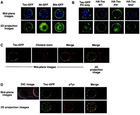
FIG. 8.
Tec, but not Itk or Btk, is localized in clusters at the cell membrane in Jurkat cells. (A) Jurkat cells were transfected with GFP-tagged Tec, Itk, or Btk. Twenty hours posttransfection, cells were fixed on glass slides and imaged by florescence microscopy with deconvolution software. Representative cells are presented with GFP in green and nuclear staining in blue. The mid-plane image represents a single, central z-section. The two-dimensional (2D) projection represents the sum total of approximately 20 z-stacks collapsed to form one image. (B) Jurkat cells were transfected with GFP-tagged Tec PH domain or HA-tagged Tec wild type, PH, or SH2 point mutants. For detection of HA-tagged Tec, cells were fixed and permeabilized and then stained with Alexa 488-conjugated anti-HA MAb (shown in green). Nuclear staining is shown in blue. Images were collected as described for panel A. (C) Jurkat cells were transfected with GFP-tagged Tec. For detection of GEMs, cells were fixed and stained with biotinylated cholera toxin followed by Alexa 647-conjugated streptavidin (shown in red). Images were collected as described for panel A. (D) Jurkat cells were transfected with GFP-tagged Tec. For detection of phosphotyrosine, cells were fixed and permeabilized and then were stained with antiphosphotyrosine MAb 4G10 followed by Cy5-conjugated antimouse antibody (shown in red). Images were collected as described for panel A. The DIC image is shown to indicate the absence of a phosphotyrosine signal in nontransfected cells.

