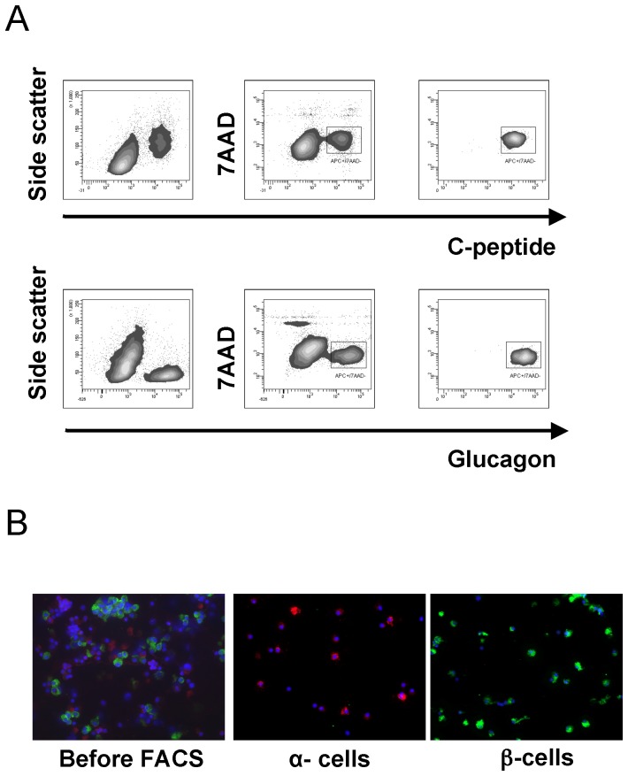
Figure 1. Intracellular staining and Flow-cytometric sorting of β-cells and α-cells.
(A) c-peptide+/7AAD− and glucagon+/7AAD− dissociated human islet cells were isolated by FACS. The right panel shows the flow cytometric analysis of the isolated populations. (B) Immunostaining of islet cells before and after FACS. C-peptide positive cells are shown in green and glucagon expressing cells in red.

