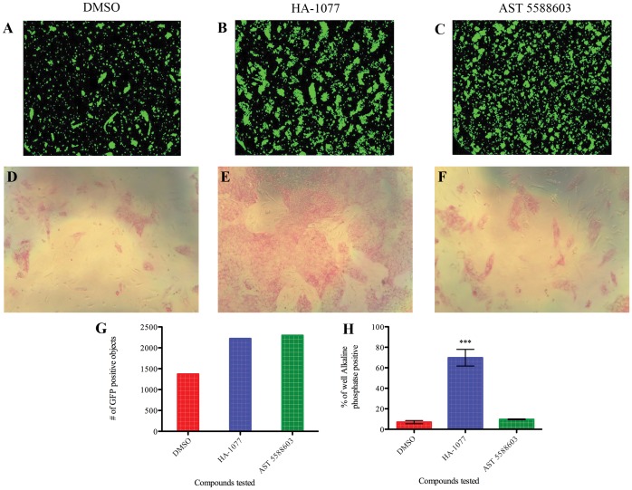
Figure 2. Small molecules identified in the OCT4-GFP screening assay were not confirmed in secondary assays.
Three treatments are compared to assess changes in survival of dissociated hESCs: 0.1% DMSO (negative control; A,D), 10 µM HA-1077 (positive control; B,E), and 10 µM AST 5588603 (representative of small molecules identified in OCT4-GFP screening assay; C,F). A-C: Readout from the Acumen microplate cytometer. The wells treated with HA-1077 and AST 5588603 had similar numbers of GFP positive objects (quantified in G). D-F: Alkaline phosphatase staining of cells treated as described. Pluripotent cells are stained with a red color. Wells treated with HA-1077 (E) contain significantly more pluripotent cells than cells treated with DMSO or any other candidate compounds. G: Quantification of GFP signal detected in (A-C). H: Quantification of alkaline phosphatase staining in (D-F) by calculating what percent of the well area stained positive for alkaline phosphatase. Data shown as mean ±SD, ***: p<0.001.

