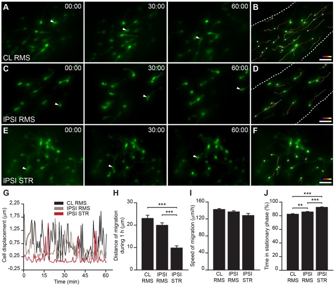
Figure 5. Dynamic properties of neuroblast migration in ischemic areas.
A–F, Timeline snapshots from real-time images of SVZ-derived precursor migration in acute brain slices of MCAo-challenged animals. Cells were labeled by injecting GFP-encoding retroviruses in the SVZ prior to inducing ischemia and were evaluated 2 to 3 weeks post-MCAo in the contralateral RMS (A,B), ipsilateral RMS (C,D), and ipsilateral striatum (E,F). Arrowheads indicate migrating cells. B,D,F, Migratory tracks of the cells migrating for 1 h in A,C,E, respectively (color code: blue in the initial position; white in the final position). G, Representative profile of cell displacement over time for individual neuroblasts migrating in the contralateral and ipsilateral RMS and the striatum. H , J, In the ipsilateral striatum, the recruited neuronal precursors displayed low migratory behavior, spending longer periods in the resting state (J), which resulted in shorter displacements per hour compared to the contralateral or ipsilateral RMS (H). I, No differences in the speed of migration of neuroblasts migrating in the ischemic striatum and ipsilateral and contralateral RMS were observed. Scale bars: 20 µm (CL: contralateral; IPSI: ipsilateral; STR: striatum).

