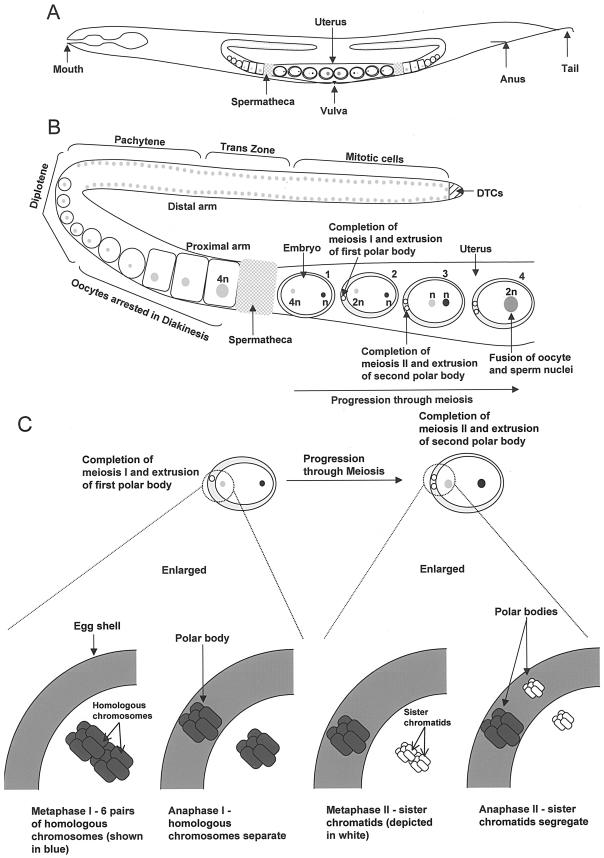
FIG.2.
(A) Adult hermaphrodite showing mouth, anus, and tail with U-shaped gonad. The spermatheca, uterus containing embryos, and vulva are also highlighted (more details in text). (B) Magnified view of one-half of the U-shaped gonad from the adult hermaphrodite with oocyte chromosomes in newly fertilized embryos undergoing meiosis in the uterus (adapted from reference 57). Embryo 1, fertilization of oocyte (grey circle, 4n) by the sperm; embryo 2, completion of meiosis I by oocyte chromosomes (grey circle, 2n) and extrusion of the first polar body; embryo 3, completion of meiosis II and extrusion of the second polar body; embryo 4, fusion of oocyte and sperm nuclei (see the text for more description). (C) Chromosome segregation during meiosis in the C. elegans oocyte (57). Six pairs of homologous chromosomes undergo meiosis I upon fertilization, followed by sister chromatid separation during meiosis II (see the text for details).

