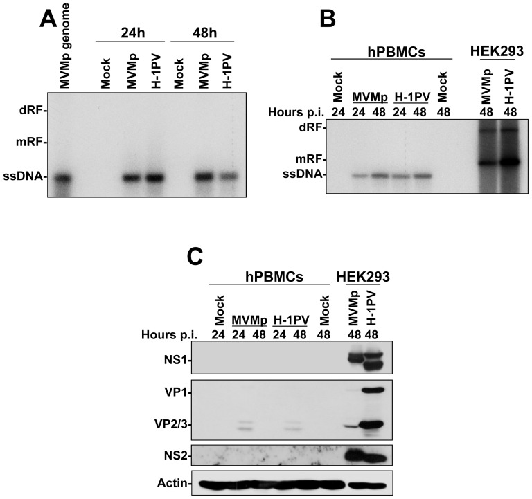
Figure 4. Defective replication of parvoviruses in hPBMCs.
(A) hPBMCs collected from the blood of healthy donors were distributed into 6-well plates at 1×10⁷ cells/5 ml culture medium/well. They were then mock-treated or infected with MVMp or H-1PV at 10 PFUs/cell and 24 hrs later harvested for Southern blotting as described in Figure 2. The presented blot is representative of 3 additional which gave similar results. (B) hPBMCs (1×10⁷ cells/5 ml culture medium/well of a 6-well plate) and HEK293 (1.5×10⁶ cells/10-cm dish) were mock-treated or infected with the indicated parvovirus at 10 and 2 PFUs/cell respectively, and for the indicated period of time. Cultures were then harvested for Southern blotting as described in Figure 2. The presented blot is representative of 2 additional which gave similar results. (C) Expression of the parvovirus proteins in infected hPBMCs was examined in the same samples as those already analyzed for parvovirus replication in (B). At the time point indicated mock-treated or parvovirus-infected cultures were harvested by scraping in PBS and centrifuged. Cell pellets were then re-suspended in complete Ripa buffer supplemented with phosphatase and protease inhibitors. Total proteins were extracted from each sample as described in Materials and Methods. Fifty µg total proteins per sample were then subjected to 8% SDS-PAGE, transferred onto membranes, and probed with laboratory produced polyclonal antibodies specific for parvovirus NS1, NS2 and capsid polypeptides (Note: The antibody specific to VP proteins was designed to detect H-1PV polypeptides and is therefore less specific for MVMp). Actin was used as an internal loading control. The presented blot is representative of 2 additional which gave similar results.

