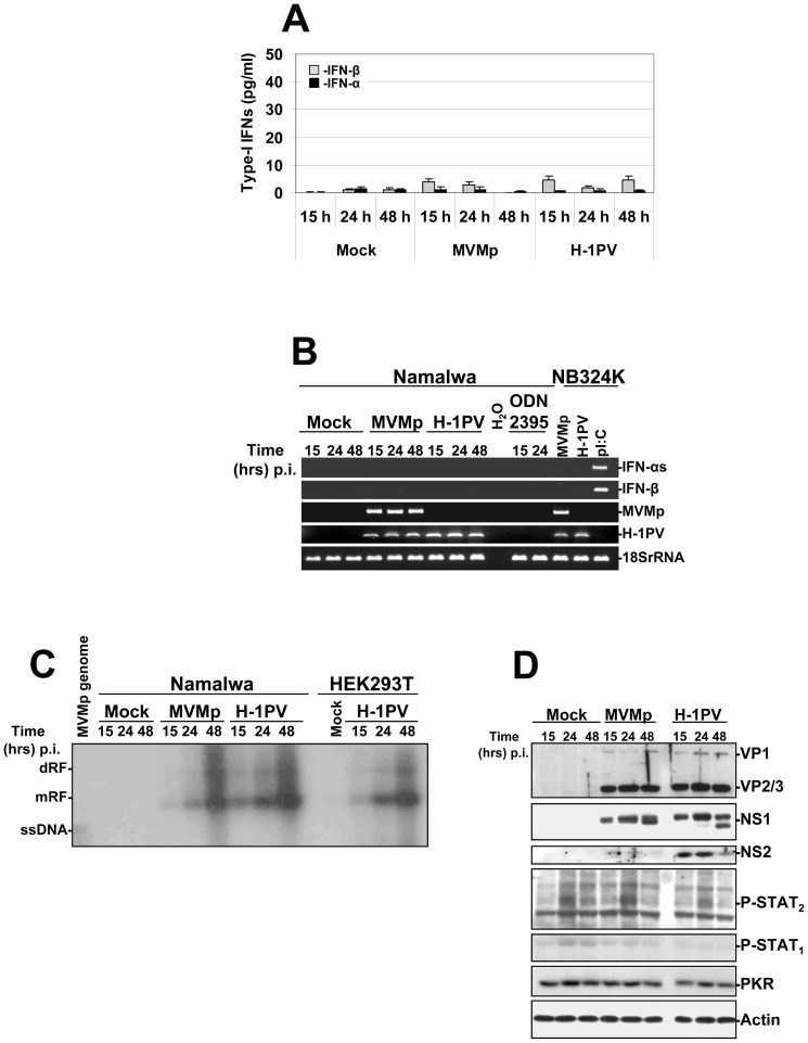
Figure 11. Activation of a TLR-9 dependent production of type-I IFNs in parvovirus-infected human Namalwa cells.
The cells (1.106 cells/well of a 24-well plate) were either infected with MVMp or H-1PV (∼80 PFUs/cell equivalent to 1×10⁴ virus genomes/cell), stimulated with the TLR-9 agonist ODN 2395 at 1 µM, or mock-treated. (A) At the indicated time points culture supernatants were collected from the respective wells and used to determine the quantity of type-I IFNs released in the culture medium by ELISA experiments. Results are expressed as means+standard deviations of three independent experiments. (B) Mock-treated, parvovirus-infected or ODN stimulated Namalwa cells were harvested at the indicated time points and total RNAs were extracted using the RNeasy kit as described previously in Figure 1. Total RNAs extracted from parvovirus-infected (24 hrs p.i. at 80 PFUs/cell) or pI:C-transfected (15 hrs post-transfection with 2 µg dsRNA/ml) NB324K cells were used as positive or negative controls. Presented data are representative of 3 experiments which all gave similar results. (C) Total DNA was harvested at the indicated time points from parvovirus-infected (MOI of 80 PFUs/cell) or mock-treated Namalwa or HEK293T cultures to assess by Southern blot experiments as described in Figure 2 the replication of both parvoviruses (dRF, dimmer replicative form; mRF, monomeric replicative form; ssDNA, single-stranded DNA genome). The blot shown is representative of 3 experiments which gave similar results. (D) Cell pellets from mock-treated or parvovirus-infected Namalwa cultures were re-suspended in complete Ripa buffer and Western blotting was performed as described in Figure 6. Actin was used as an internal loading control. Each presented blot is representative of 3 experiments which gave similar results.

