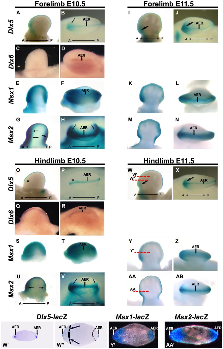
Figure 1. Spatio-temporal expression of Dlx-Msx in developing limbs.
A-N, forelimbs. O-AB, hindlimbs, at E10.5 (A-H, O-V) and E11.5 (I-N, W-AB). Whole-mount X-gal staining on FLs and HLs from Dlx5+/−, Msx1+/− and Msx2+/− (lacZ+) heterozygous embryos are shown. Expression of Dlx6 was detected by WMISH on embryonic limbs at the same ages and shown. At E10.5 Msx2 and Msx1 are expressed in the AER and the anterior and posterior mesoderm of HLs and FLs. Dlx5 and Dlx6, at E10.5, are expressed in the AER of HLs and FLs and in the anterior limb mesoderm only of the FLs, but not of the HLs. At later stages (E11.5), Dlx5 and Dlx6 are then expressed in the anterior mesoderm of HLs. Black arrows indicate mesodermal expression. The AER is also indicated. Black asterisks indicate absence of expression. W’,W’’ histologic transversal sections of E11.5 HLs from Dlx5+/− embryos, stained with Xgal. Y’,AA’ histologic transversal sections of HLs from Msx1+/− (Y’) and Msx2+/− (AA’) embryos, to compare AER and mesodermal expression between these genes. Section planes and position are reported with red lines (in W, Y and AA). The extent of the Msx1-positive anterior and posterior mesoderm regions, based on the micrographs in AA’ and AC’, are indicated with dashed lines. A strong Dlx5-lacZ signal is detected in the AER (W’ and W’’), a weak Dlx5-lacZ signal, overlapping with the Msx1-lacZ and the Msx2-lacZ signal, is detected in the anterior mesoderm (W’’, indicated by black arrows).

