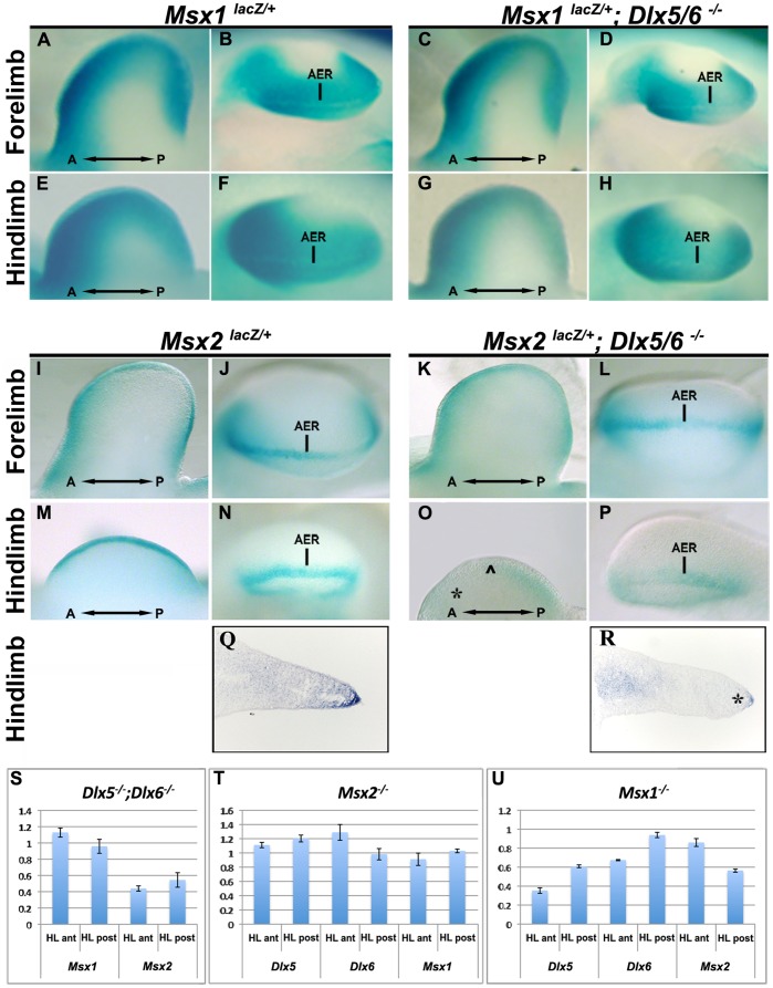
Figure 2. Reduction of Msx2 expression in Dlx5/Dlx6 DKO HLs.
A-P. Whole-mount X-gal staining to detect Msx1 and Msx2 expression in Dlx5;Dlx6 DKO. In the FL (A-D,I-L) no changes of expression is observed whereas in the HL (E-H, M-P), Msx2 expression is reduced in the AER and in the anterior limb mesoderm of the Dlx5;Dlx6 DKO HLs. Q,R. Sections of WT (Q) and Dlx5;Dlx6 DKO mutant HLs (R) hybridized in situ to detect Msx2, showing a drastically reduced Msx2 signal in the AER and in the underlying mesoderm, but not in a proximal mesoderm territory. S-U. Quantification of the expression of Dlx5, Dlx6, Msx1 and Msx2 mRNAs by qRT-PCR in HLs from Dlx5;Dlx6 DKO (S), Msx2−/− (T) and Msx1−/− (U), relative to WT. The results show a reduction of 45% of Msx2 expression in the Dlx5;Dlx6 DKO HLs compared to WT, but not of Msx1. Dlx5 and Dlx6 expression is downregulated in Msx1 KO HLs but not in Msx2 KO HLs. Expression of the knocked-out genes was also tested, as control, and always found to be reduced to undetectable levels (not shown).

