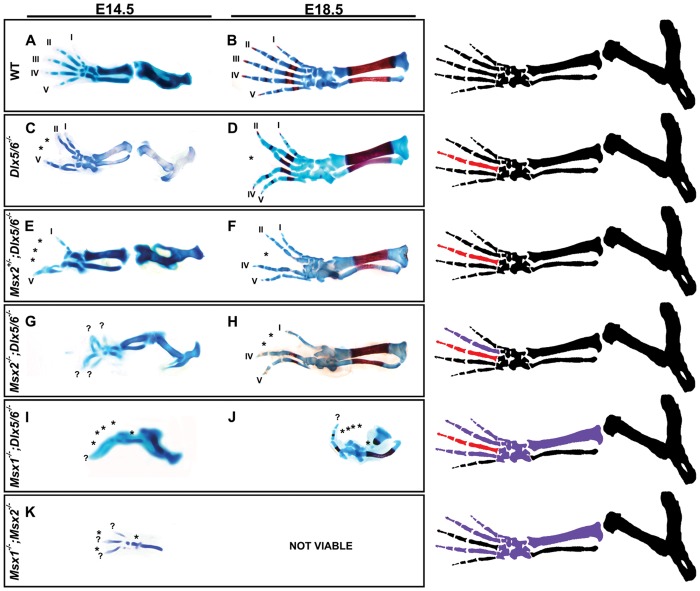
Figure 3. Skeletal preparations of the HLs of single and combined Msx;Dlx mutant animals.
Chondroskeletal preparation of the HLs of E14.5 embryos (micrographs on the left) and full skeletal preparation on newborn animals (micrographs on the right), representing single and combined Dlx;Msx mutant genotypes (indicated on the left). The HLs of Msx2 +/−;Dlx5;Dlx6 DKO animals (E,F) display an aggravated ectrodactyly phenotype compared to Dlx5;Dlx6 DKO ones (C,D), with fusion of the external digit (1with 2, 4 with 5) and hypoplasia of the central digit. Msx2;Dlx5;Dlx6 TKO HLs (G,H) display a further aggravated ectrodactyly phenotype, with the external digits fused and extended towards the opposite (anterior-posterior) sides, and a complete absence of the central digit. The limbs of Msx1+/−;Dlx5;Dlx6 DKO mice (not shown) show ectrodactyly similar to that observed in Dlx5;Dlx6 DKO mice, whereas Msx1;Dlx5;Dlx6 TKO mice (I,J) show ectrodactyly and loss of skeletal elements deriving from the anterior mesoderm of the autopod and zeugopod, a phenotype seen in Msx1;Msx2 DKO mutant embryos (K). The stylopod shows no evident defects. The anterior-posterior orientation is shown. The numbers 1–5 indicate the digits (1 is the toe). Asterisks indicate hypoplasia or absence of skeletal structures. The drawings on the left schematically illustrate the Dlx-related (red elements) and the Msx-related (purple elements) skeletal defects, corresponding to the genotypes examined.

