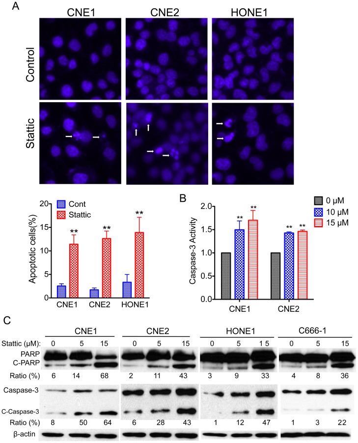
Figure 4. Stattic induces apoptosis in NPC cells.
(A) Apoptosis was measured by Hoechst 33342 staining. (Top) NPC cells were treated with 10 µM Stattic for 48 h, nuclei were stained with Hoechst 33342, and imaging analysis was performed as described in the Materials and Methods. The white arrows indicate apoptotic cells. Original magnification, ×200. (Bottom) Quantification of the cell staining. (B) Effect of Stattic on caspase-3 activity. The cells were treated with the indicated concentrations of Stattic for 48 h. The activities were determined as described in Materials and Methods. (C) NPC cells were exposed to the indicated concentrations of Stattic for 48 h; apoptotic cells were measured by western blot analysis of cleaved PARP and cleaved caspase-3. Protein levels were quantified using ImageJ software. Data are means ± s.d. for three independent experiments, *P<0.05, **P<0.01. DMSO were used as control in “0” groups.

