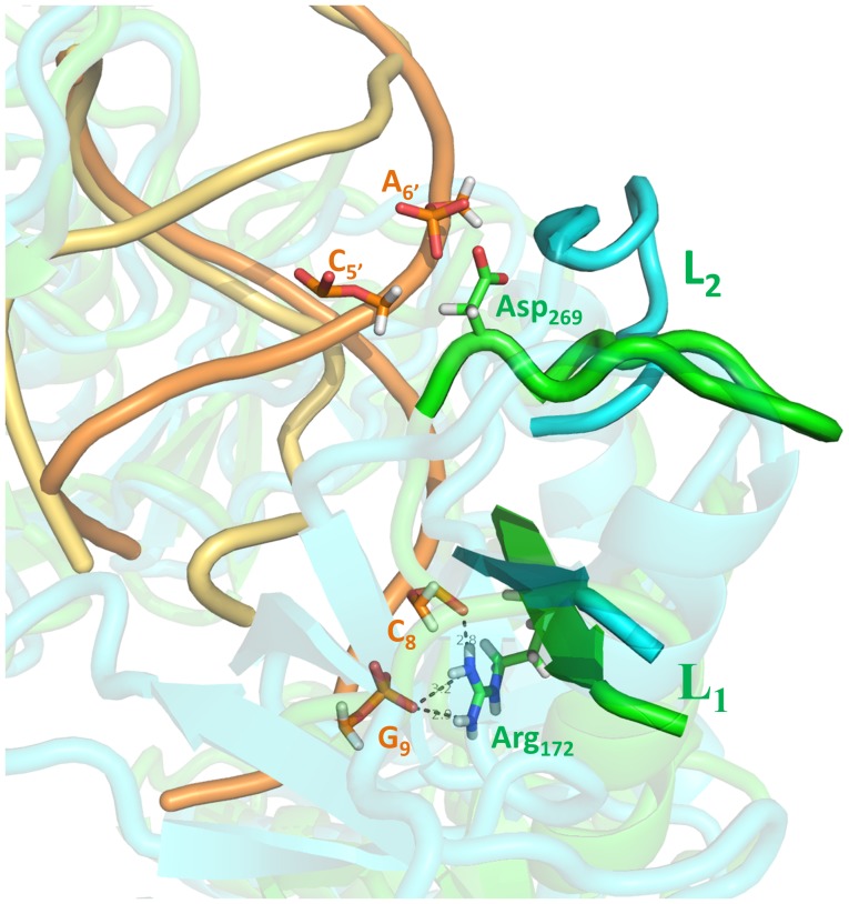
Figure 7. A closer view of the “hinge-like” bending at L1/L2 segment by superposing the final conformation and the starting native structure of the 4-site mutant with 11-bp guide DNA-target RNA duplex.
The nucleic acid and L1/L2 segment in the wild-type are colored orange and green, respectively. The nucleic acid in the 4-site mutant is colored yellow and the L1/L2 segment is colored cyan. The residues that form hydrogen bonds between the guide-target duplex and L1/L2 segment are shown as sticks.

