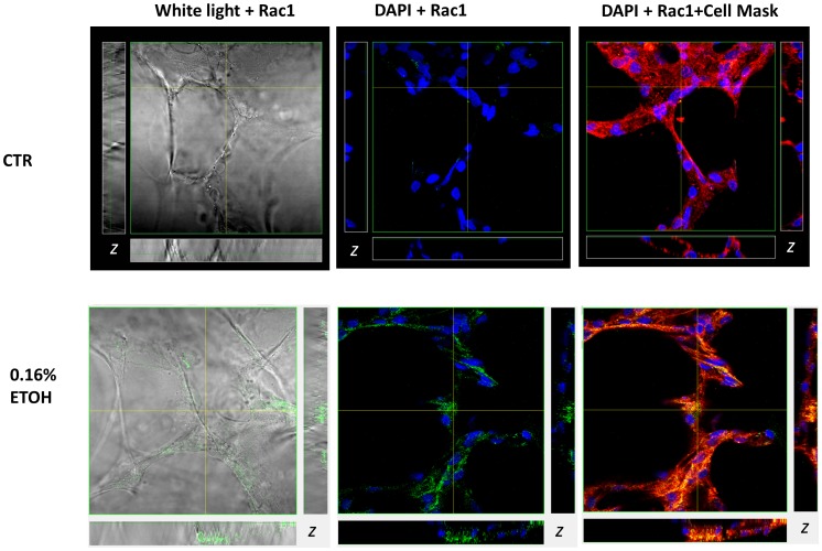
Figure 8. Multichannel confocal imaging shows EtOH mediated changes in Rac1 expression and subcellular localization in the lung.
Anti-Rac1 antibody detected using Alexa 488 conjugated secondary antibody (488/519 nm; green fluorescence); nuclei were stained DAPI (350/470 nm; blue fluorescence); and plasma membrane labeled with Cell Mask Deep Red (649/666 nm). Right panel: white light image of lung slice preparation and Rac1 labeling; middle panel: Rac1 localization relative to DAPI stained nuclei; left panel: Rac1 co-localization with Deep Red labeled plasma membrane. Pixels containing both red and green color contributions produce various shades of orange and yellow indicate Rac1 co-localization with the plasma membrane. Subsets represent z-axis obtained from horizontal and vertical regions as indicated.

