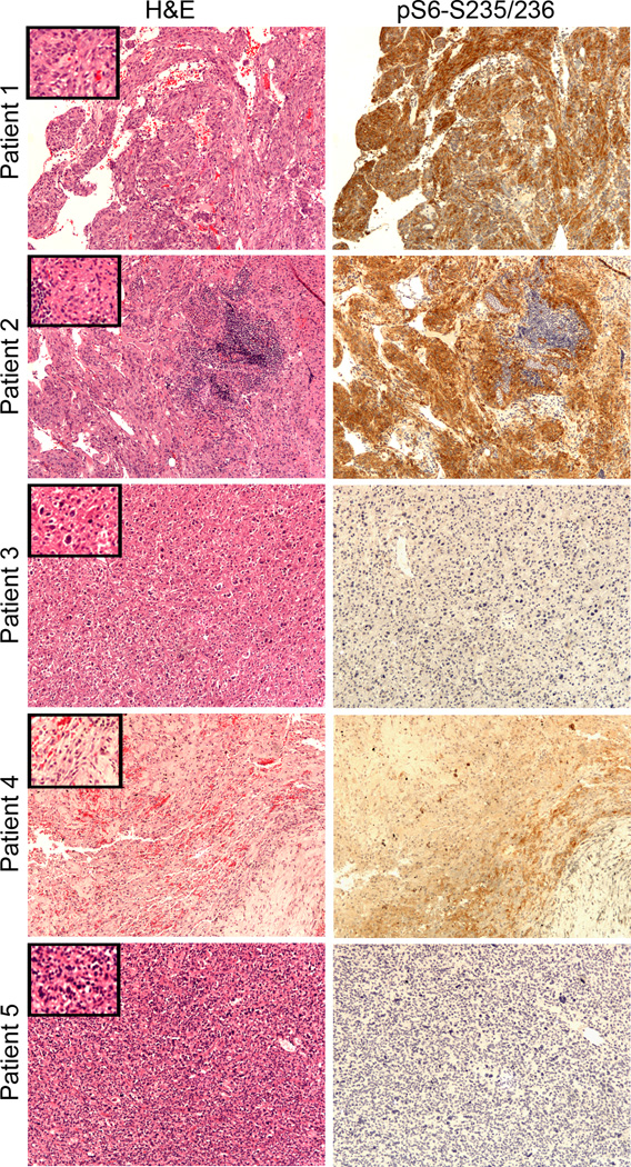
Figure 2.
Pathology and immunohistochemistry analysis of PEComas. H&E staining and immunostaining for phospho-S6 protein (Ser235/236) in all tumors samples (10× objective; insets-20×). PEComas from patients 1 and 2 are strongly pS6 positive, whereas the PEComas of patient 3, 4 and 5 are not. In patient 2 normal lymphoid tissue is negative for pS6-S235/236. In Patient 4 areas of hemorrhage, hemosiderin deposits and variable pS6 staining are apparent, following drug treatment.

