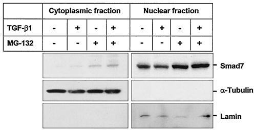
FIG. 4.
Smad7 is constitutively present in the nucleus, and TGF-β1 treatment increases Smad7 in the cytoplasm. HaCaT cells treated or not treated with 5 ng of TGF-β1/ml for 14 h were incubated in the presence or absence of MG-132, an inhibitor of proteosom-mediated degradation, for 2 h before lysis. Lysates were separated into nuclear and cytoplasmic fractions and immunoblotted with antibodies against Smad7 (top panel) and, to confirm the purity of the fractions, α-Tubulin (middle panel) and Lamin (lower panel). All three blots were prepared from the same lysate and are representative of the results obtained in three separate experiments.

