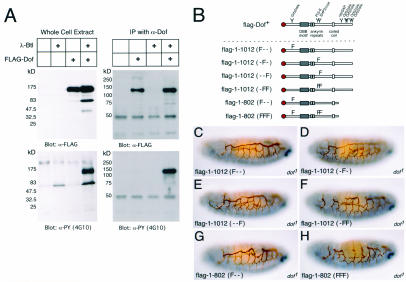
FIG. 6.
Dof phosphorylation in response to FGF receptor activation and the effect of specific tyrosine mutations on the function of Dof. (A) Drosophila Schneider S2 cell lines were stably transformed as indicated above each lane with different combinations of an activated form of the FGF receptor Btl under the control of a heat shock promoter (λ-Btl) and Dof with an N-terminal FLAG epitope tag under the control of the actin5C promoter (FLAG-Dof). The presence of Dof and proteins with phosphorylated tyrosine residues in whole-cell lysates of these cell lines, previously induced to express the FGF receptor (left), or immunoprecipitates (IP) of the induced lysates with an antiserum directed against Dof (α-Dof) (right) was monitored by Western blotting with antibodies directed against the FLAG epitope tag (M5) (α-FLAG) and phosphotyrosine (4G10) (α-PY). The sizes of the proteins detected are indicated on the left. Dof is seen at an apparent molecular mass of ∼130 kDa. Often, an additional band representing a breakdown product is seen at ∼70 kDa. (B) Schematic representations of Dof constructs. The name of a signaling protein and a Y indicates the position of a tyrosine in Dof that is a potential binding site for this molecule. An F indicates a specific mutation that results in a protein containing a phenylalanine residue rather than a tyrosine; different combinations of mutations in amino acids 97, 486, and 515 are shown. (C to H) Tracheal development of homozygous dof mutant embryos expressing the constructs flag-Dof <Y95F> (C), flag-Dof <Y486F> (D), flag-Dof <Y515F> (E), flag-Dof <Y486F, Y515F> (F), flag-Dof[1-802] <Y97F> (G), and flag-Dof[1-802] <Y97F, Y486F, Y515F> (H). The embryos are stained for 2A12 in brown and Even-skipped in blue to distinguish the tracheae and the genotype, respectively, as described in the legend to Fig. 2.

