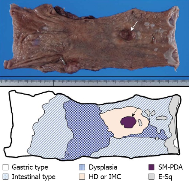
Figure 1.

Macroscopic findings of excised esophagus. Surgical specimen shows the presence of superficial spreading carcinoma, extending between 30 mm from the oral and 105 mm from the anal surgical margins. The superficial spreading region is mostly composed of high-grade dysplasia (HD), or intramucosal adenocarcinoma (IMC), surrounded by dysplastic change (E-sq low-grade dysplasia). Inside this superficial spreading region, one observable elevated nodule (arrow) is composed of solid and submucosal invasive poorly-differentiated invasive adenocarcinoma (SM-PDA) with lymphatic invasion. The background non-neoplastic esophageal mucosa is extensively replaced by glandular mucosa with and without intestinal metaplasia.
