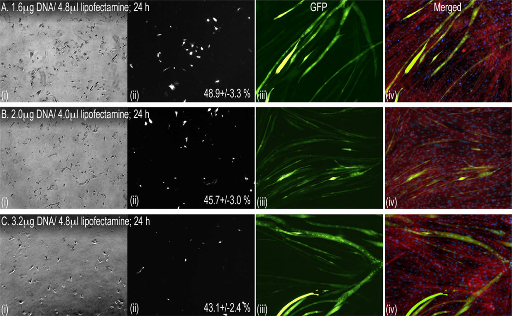
Figure 1. Optimizing transfection of C2C12 myoblasts.
Subconfluent cells were passed 24 h before transfecting with the indicated DNA/Lipofectamine ratios using the manufacturer’s protocol. Ratios above and below those indicated were either toxic or were significantly less efficient (see Results). Myoblasts were imaged 24 h (40×) after transfection, grown to confluency for 4 additional days and differentiated with serum-withdrawal (iii & iv). Shown in each panel set (A–C) are phase-contrast (i) and fluorescent images of corresponding myoblasts (ii), differentiated myotubes (iii, GFP, 100×) and merged images (iv) of GFP, DAPI-stained nuclei and sarcomeric actin labeled with phalloidin red. Transfection efficiency (mean % +/− SEM; no significant differences) is indicated for each treatment and was determined by counting fluorescent and total cell numbers in 9 random views from each well/treatment.

