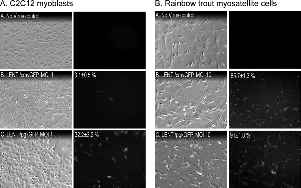
Figure 5. Comparison of pgk and cmv promoters.
(A) C2C12 myoblasts and (B) primary trout myosatellite cells were incubated for 72 h with lentivirus (LENTI) containing either cmv/GFP or pgk/GFP at the indicated MOI. Transduction efficiency (mean % +/− SEM; values in C2C12 experiments were significantly different, p≤0.05) was determined by counting fluorescent and total cell numbers in 9 random views of each well/treatment.

