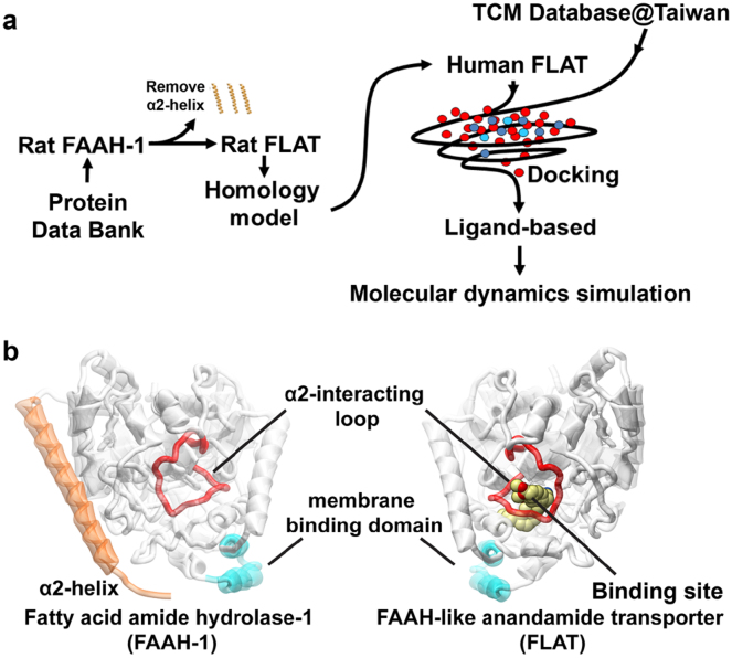
Figure 1. Experimental procedure and structural basis of FLAT simulation.
(a) Simplified scheme of experimental procedures. (b) Structural basis for FLAT structure simulation using FAAH-1. The α2-interacting loop (K255-L278; red) is the binding site opening loop, and the helices (P411-N435) colored in cyan are regions in FAAH-1 that interact with the membrane. Presence of the α2-helix (T9-T76; orange) in FAAH-1 was the primary structural difference from FLAT. Human FLAT was modeled from rat FLAT structure, which was computationally prepared by deleting the α2-helix region (amino acids T9-T76) in rat FAAH-1.

