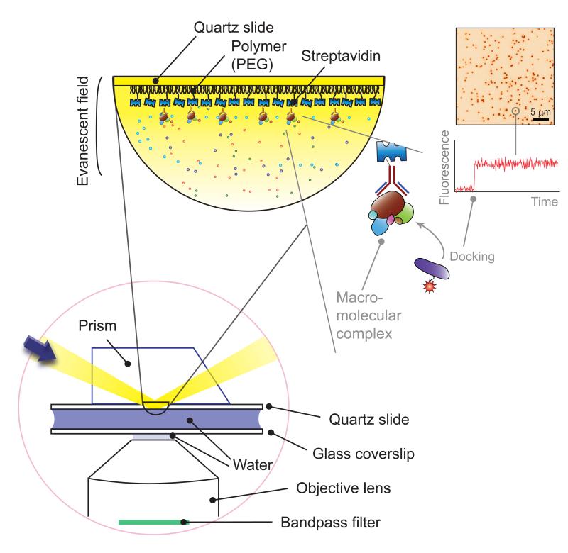
Figure 1. Total internal reflection microscopy.
(Bottom left) In total internal reflection microscopy, a sheer layer of ~100 nm on a glass surface is excited via total internal reflection at the interface of water and glass (here, quartz). Background signals from the solution are thereby effectively minimized, which is essential for harvesting a finite number of photons from single fluorophores. As the excitation is confined to the glass surface, the molecules of interest are immobilized on a surface for long-term observation. (Top left) To immobilize molecules of interest, biotin-streptavidin-biotin conjugation is used. Here, streptavidin is bound to a biotinylated polymer (PEG, poly-ethylene glycol) surface, the streptavidin proteins bind to biotinylated antibodies, and the macromolecular protein complexes are bound to the antibodies. (Top right) In this immobilization scheme, the docking of a fluorescent partner molecule leads to a sudden increase in fluorescence signals over a localized spot, as shown in a representative CCD image and fluorescence time trace. Adapted from Yeom et al. [16] with permission.

