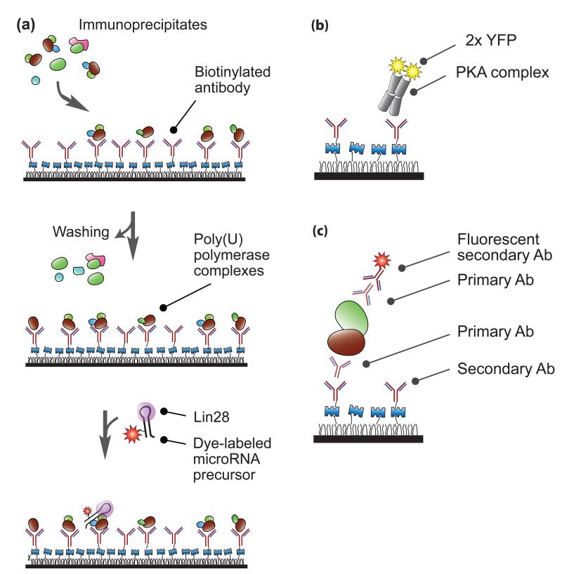
Figure 4. Single-molecule immunoprecipitation.
(a) MicroRNA modification. Using surface-bound antibodies, Yeom et al. immobilized poly(U) polymerase immunoprecipitates obtained from human cell extracts[16]. After washing to remove unbound immunoprecipitates, they introduced fluorescently tagged Lin28-bound precursor microRNA. By recording fluorescent signals from the labeled RNA substrates, they visualized interactions between the RNA strands and the protein complexes in real time. (b-c) Single-molecule complex analysis. (b) Jain et al. pulled down a protein kinase A complex using surface-immobilized antibodies[17]. The stoichiometry of the complex was analyzed based on the number of YFP (yellow fluorescent protein) photobleaching steps. (c) Endogenous protein complexes were pulled down and visualized via the combination of primary antibodies and secondary antibodies. “Ab” stands for antibody. A quartz slide is coated with polymer and layered with Streptavidin as shown in Figure 1.

