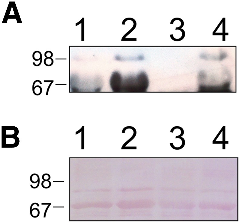
Fig. 12.

Western blot using mAb-HSA-VX of plasma samples from rats treated with VX (0.8 × LD50) for 72 hours. (A) Lanes 1 and 3, plasma (1 μl) from untreated rats; lanes 2 and 4, plasma (1 μl) from treated rats. (B) Ponceau staining of an identical gel as blot A run in parallel that showed equal protein loading of serum albumin.
