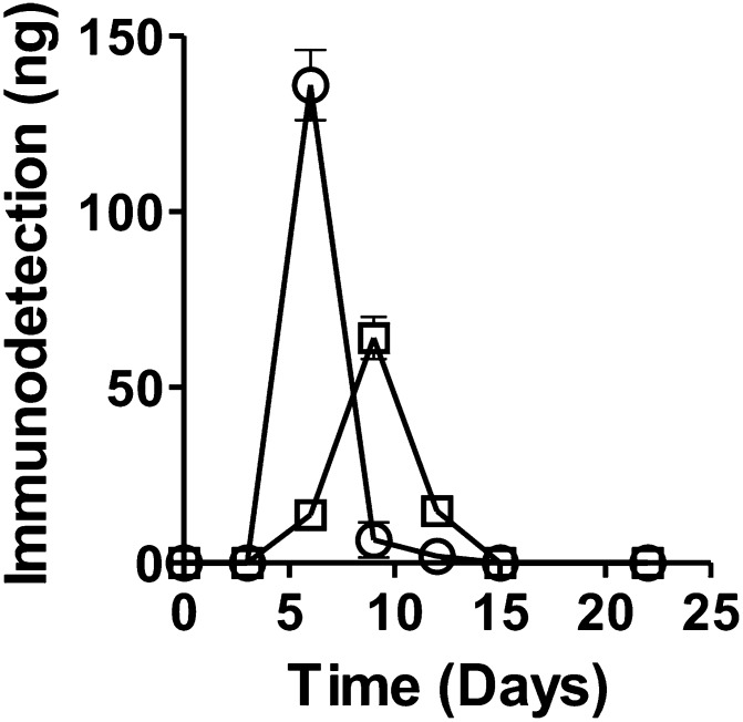
Fig. 9.
A plot of the amount of GD nerve agent model compound stereoisomers adducted to HSA as a function of time based on quantification of immunoreactivity with mAb-HSA-GD. The amount of HSA-GD protein adduct was determined by Western blot analysis. After addition of nerve agent model compound Sp-GD or Rp-GD methylthiocholine to HSA, the incubation was sampled over time and compared with an HSA-GD-adducted protein standard. Quantification was done by densitometry. ○, Sp-GD methylthiocholine; □, Rp-GD methylthiocholine.

