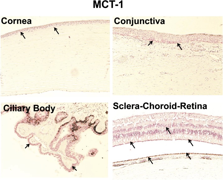
Fig. 8.
Representative figure of immunohistochemical localization of MCT-1 in human ocular tissues. Arrowhead indicates the localization of MCT-1 staining. MCT-1 showed light expression in corneal and conjunctival basal epithelial cells. MCT-1 showed strong labeling in the nonpigmented ciliary epithelium compared with any other ocular tissues. For choroid-retina, MCT-1 staining was present in the outer segment of photoreceptor cells, inner limiting membrane, and RPE cell layer. Nuclei were counterstained in blue with hematoxylin.

