
Figure 2.
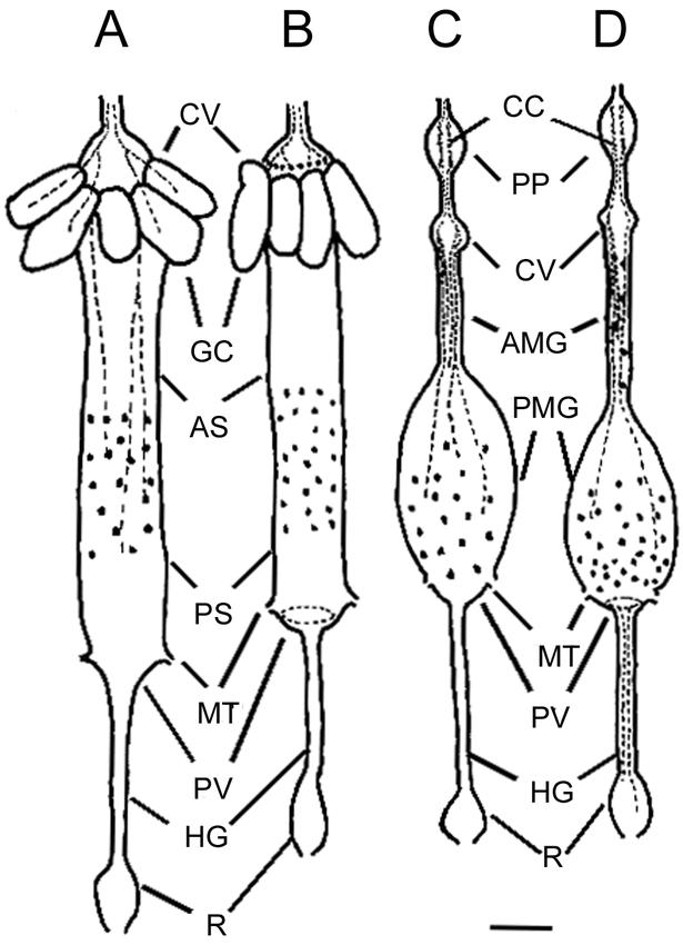
Distribution of OEH I immunostained cells and axons in the stomatogastric nervous system and gut of fourth instar larvae of Aedes aegypti, A, and Anopheles gambiae, B, and female Ae. aegypti, C, and An. gambiae, D. AMG, anterior midgut; AS, anterior stomach; CC, corpus cardiacum; CV, cardiac valve; GC, gastric caeca; HG, hindgut; MT, Malpighian tubules; PMG, posterior midgut; PS, posterior stomach; PV, pyloric valve; R, rectum. Filled circles: immunostained cells, dotted lines: immunostained axons. Scale bar = 400 µm.