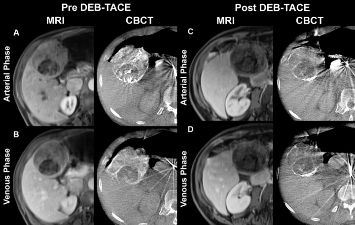
Figure 2:
Representative T1-weighted axial contrast-enhanced arterial and portal venous phase MR images (MRI) and axial contrast-enhanced early or arterial phase and delayed or venous phase cone-beam CT scans (CBCT) in 73-year-old man with cryptogenic HCC in right lobe. MR images were acquired approximately 1 month before and 1 month after TACE with doxorubicin-eluting beads (DEB), whereas dual-phase cone-beam CT scans were acquired intraprocedurally before and immediately after delivery of doxorubicin-eluting beads during TACE. A, Arterial phase images obtained before TACE. MR image shows a 75-mm mass in right lobe with 65% enhancement. Mass has similar enhancement (75%) on cone-beam CT scan. B, Venous phase images obtained before TACE. MR image shows mass with 60% enhancement. Similar enhancement (70%) is seen on cone-beam CT scan. C, Arterial phase images obtained after TACE. Mass shows almost no enhancement (5%) on MR image, and tumor has decreased to 69 mm. On cone-beam CT scan, intraprocedural mass enhancement decreased by 87%, which allowed prediction of an objective EASL response at 1 month. D, Venous phase images obtained after TACE. Mass shows no enhancement (0%) on MR image. Intraprocedural enhancement decreased by 93% on cone-beam CT scan, enabling prediction of an objective EASL response at 1 month. As shown here, the patient has shifted in position between acquisition of the post-TACE MR images and the cone-beam CT images and pre-TACE MR images, which explains some differences in tumor size and morphology despite the fact that images were obtained at same level.

