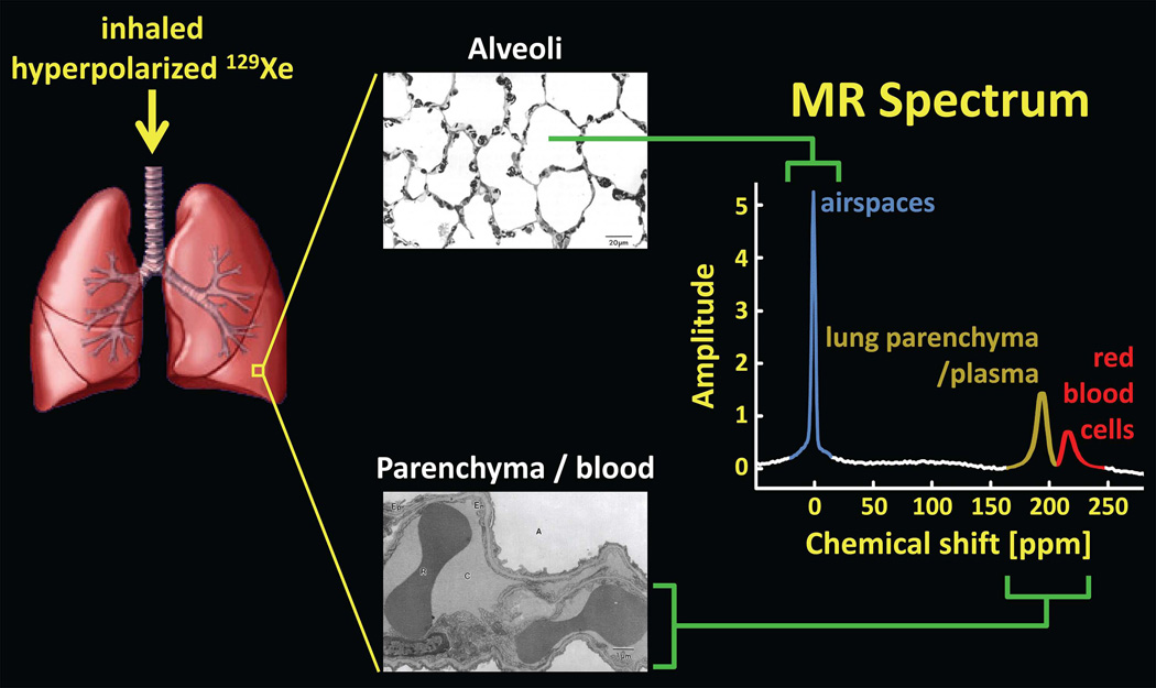
Figure 10.
129Xe in the lung. Inhaled xenon gas distributes among the airspaces of the lung and also dissolves in the lung parenchyma and blood. Because of the large chemical-shift, and hence frequency, difference between 129Xe in the airspaces (alveoli) and that in the parenchyma/blood, an MR spectrum of 129Xe in the lung reveals distinct spectral peaks corresponding to the gas and dissolved components. For clarity, the dissolved-phase peaks, associated with roughly 2% of 129Xe in the lung, are shown several times larger than they would appear if the same flip angle was applied to both the gas-phase and dissolved-phase compartments. (Lung rendering adapted from: dir.niehs.nih.gov/dirlrb/mcb/proj-cox.htm. Micrographs reproduced with permission from Albertine KH. Structural organization and quantitative morphology of the lung. In: Cutillo AG, ed. Application of Magnetic Resonance to the Study of Lung. Hoboken, NJ: Wiley-Blackwell, 1996; 73–114.)

